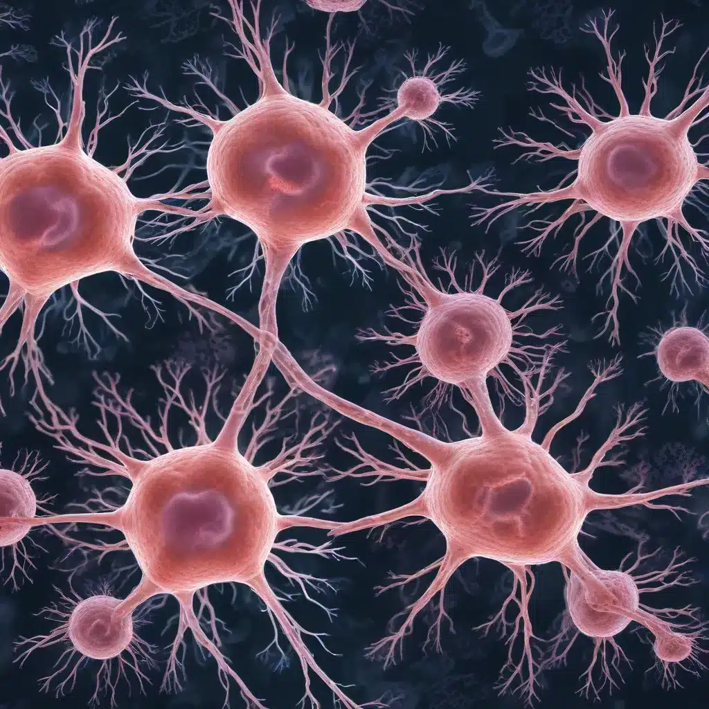
Obesity, Neurodegenerative Disorders, and the Adipocyte-Brain Axis
Obesity and type 2 diabetes are well-established risk factors for neurodegenerative disorders like Alzheimer’s and Parkinson’s disease. While the exact mechanisms linking these conditions remain poorly understood, emerging research suggests that disruptions in the communication between adipose tissue (fat) and the brain play a crucial role.
Adipose tissue is no longer viewed as a passive energy storage depot. Instead, it is recognized as a dynamic endocrine organ that secretes a variety of signaling molecules, including lipids and adipokines, which can directly influence brain function and neuronal health. This adipocyte-brain axis is essential for regulating feeding behavior, energy expenditure, and even cognitive processes.
Interestingly, when this intricate communication between adipose tissue and the brain is disrupted, it can contribute to the development of neurodegenerative disorders. One key mechanism by which this may occur is through the impact of adipocyte metabolism on the function of glial cells in the brain.
Glial Cells: The Brain’s Cleanup Crew
Glial cells, often referred to as the “cleanup crew” of the brain, perform essential functions in maintaining a healthy neural environment. These cells, which include microglia and their Drosophila counterparts, the ensheathing glia, are responsible for clearing away cellular debris and protein aggregates that accumulate with age and injury.
By phagocytosing (engulfing and digesting) this neural waste, glial cells help prevent the buildup of toxic materials that can lead to secondary neuronal death and neurodegeneration. The ability of glial cells to efficiently carry out this phagocytic function is crucial for maintaining brain health and preventing the onset of neurodegenerative diseases.
Adipocyte Metabolism and Its Impact on Glial Function
Emerging research, including studies using the Drosophila model system, has revealed a surprising and direct link between the metabolic state of adipocytes and the phagocytic function of glial cells in the brain. This connection highlights a novel pathway by which obesity and related metabolic disorders may increase the risk of neurodegenerative conditions.
Adipocyte Metabolic Adaptations to an Obesogenic Diet
When exposed to a prolonged high-sugar diet (HSD), which can induce obesity, Drosophila adipocytes undergo a metabolic shift. Instead of primarily utilizing glycolysis (the breakdown of glucose) to meet their energy needs, these cells start to rely more heavily on fatty acid oxidation (FAO) and ketogenesis (the production of ketone bodies).
This metabolic adaptation is reminiscent of the body’s starvation response, where the lack of readily available glucose forces cells to turn to alternative fuel sources, such as stored fats. Interestingly, this HSD-induced metabolic state in adipocytes is accompanied by changes in mitochondrial morphology and an increase in antioxidant defenses, likely as a protective mechanism against the potential for increased oxidative stress.
Adipocyte Metabolism Regulates Glial Phagocytic Function
The dramatic metabolic changes observed in adipocytes in response to an obesogenic diet have a surprising and profound impact on the function of glial cells in the brain. Through a series of genetic manipulations in Drosophila, the researchers have discovered that the metabolic state of adipocytes, particularly the availability of fatty acids and ketone bodies, directly regulates the expression of the phagocytic receptor Draper in ensheathing glia (the Drosophila counterpart of mammalian microglia).
Specifically, they found that increasing fatty acid availability in adipocytes, either by reducing the expression of the fatty acid transport enzyme Cpt1 or by decreasing the expression of the lipid droplet-associated protein Plin1, leads to a reduction in Draper levels in ensheathing glia. Conversely, genetically reducing the production of ketone bodies in adipocytes (by knocking down the enzyme Acat1) resulted in an increase in Draper expression.
These findings suggest that the metabolic alterations induced by an obesogenic diet, including increased fatty acid oxidation and ketogenesis in adipocytes, can negatively impact the phagocytic capacity of glial cells in the brain.
The Role of Adipocyte-Derived ApoB Lipoproteins
The researchers further identified a key signal from adipocytes that regulates glial phagocytic function: the Drosophila equivalent of human apolipoprotein B (ApoB), known as Apolpp.
ApoB is the primary protein component of lipoproteins, which are responsible for transporting lipids from peripheral tissues, including adipose tissue, to the brain. In Drosophila, Apolpp-containing lipophorins play a crucial role in delivering lipids to the brain and supporting neuronal function.
Interestingly, the researchers found that knocking down Apolpp expression specifically in adipocytes reduced the levels of the phagocytic receptor Draper in ensheathing glia, both at baseline and in response to neuronal injury. This suggests that the adipocyte-derived Apolpp signal is essential for maintaining glial phagocytic competence.
Moreover, they discovered that the low-density lipoprotein receptor (LpR1), expressed on the surface of ensheathing glia, is required for the proper upregulation of Draper in response to neuronal injury. Disrupting LpR1 function in glia impaired their ability to clear away damaged neuronal debris, highlighting the importance of the adipocyte-to-glia Apolpp-LpR1 signaling axis in supporting glial phagocytic function.
Implications for Managing Obesity-Related Cognitive Decline
The findings from this Drosophila research provide critical insights into the mechanisms by which obesity and related metabolic disorders can increase the risk of developing neurodegenerative conditions. By demonstrating that the metabolic state of adipocytes directly regulates the phagocytic capacity of glial cells in the brain, this study offers a novel perspective on the adipocyte-brain axis and its potential implications for managing obesity-related cognitive decline.
Importantly, the study identifies several potential therapeutic targets that could be explored to support glial function and mitigate the neurodegeneration associated with obesity and type 2 diabetes. These include:
- Modulating adipocyte fatty acid and ketone body metabolism
- Enhancing the Apolpp-LpR1 signaling axis between adipocytes and glia
- Supporting glial phagocytic capacity through targeted interventions
As the Stanley Park High School community continues to navigate the challenges of obesity and its impact on student health and academic performance, understanding the intricate connections between adipose tissue, glial function, and neurodegeneration may prove invaluable. By incorporating these findings into our educational and wellness initiatives, we can empower our students and families to make informed choices that support brain health and cognitive function throughout their lives.
For more information on the school’s health and wellness programs, please visit our website. If you have any questions or would like to get involved, don’t hesitate to reach out to our school nurse or wellness coordinator.

