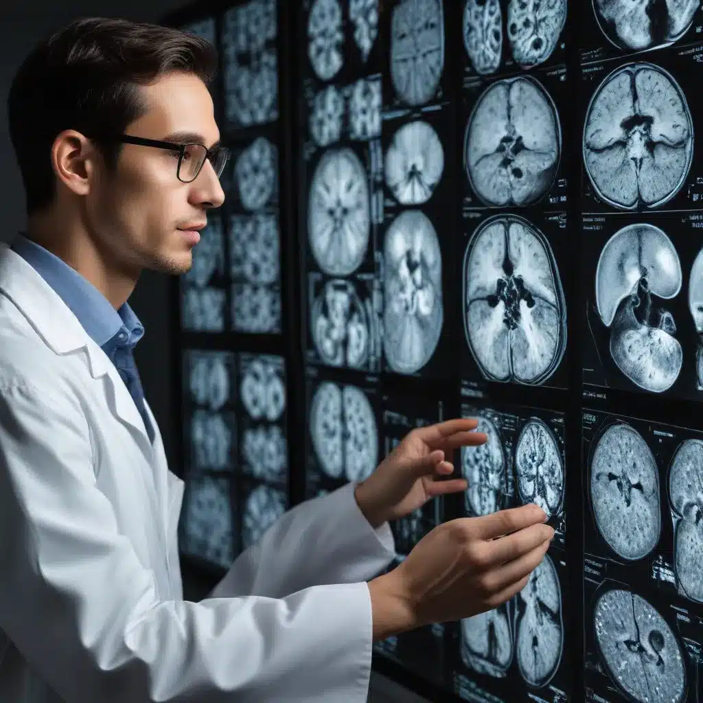
Unlocking the Secrets of the Brain: The Transformative Role of Neuroimaging
Neuroimaging has revolutionized our understanding of the brain and become an essential tool for researchers studying neurological disorders. Functional magnetic resonance imaging (fMRI) and electroencephalography (EEG) are two widely used neuroimaging techniques that provide valuable insights into brain function and activity.
fMRI is a non-invasive technique that uses magnetic fields and radio waves to produce detailed brain images, while EEG records the brain’s electrical activity through electrodes placed on the scalp. Recent advancements in these technologies have significantly expanded their applications in neuroscience research and clinical practice.
Pushing the Boundaries of fMRI
Cutting-edge fMRI technology has seen remarkable improvements in recent years. Higher spatial resolution allows researchers to study smaller brain structures and activity patterns with greater precision. Real-time fMRI enables the observation of brain activity in real-time, opening up new possibilities for therapeutic interventions such as neurofeedback. The integration of fMRI with other imaging modalities, like EEG and magnetoencephalography (MEG), provides a more comprehensive understanding of brain function by combining spatial and temporal information.
Resting-state fMRI, which measures brain activity while a person is at rest, has emerged as a powerful tool for identifying functional connectivity between brain regions and detecting changes in brain activity due to neurological disorders. Ultra-high field fMRI, operating at 7 Tesla or higher, offers increased sensitivity and spatial resolution, further enhancing our ability to study the brain in detail.
Advancements in EEG Technology
EEG technology has also experienced significant advancements in recent years. High-density electrode arrays with hundreds of electrodes allow for more precise localization of brain activity and better capture the dynamics of neural networks. Real-time source localization enables the identification of the location and strength of brain activity in real-time, which is particularly useful for applications such as neurofeedback and brain-computer interfaces.
The integration of EEG with other neuroimaging techniques, such as fMRI, provides complementary information about brain function, enhancing our understanding of the spatial and temporal characteristics of neural activity.
Unveiling the Mysteries of the Brain
Neuroimaging techniques have revolutionized our ability to study cognition, emotion, perception, and other fundamental aspects of brain function by capturing the dynamic nature of brain processes. These tools have facilitated breakthroughs in our understanding of brain organization, connectivity, and plasticity, opening new avenues for studying the neural correlates of behavioral, cognitive, and psychiatric disorders.
Neuroimaging has also played a critical role in the clinical applications of brain research. It has enabled the identification of biomarkers, assessment of treatment response, and development of personalized therapies for various neurological and psychiatric disorders, such as autism spectrum disorder (ASD), attention deficit hyperactivity disorder (ADHD), Alzheimer’s disease (AD), and Parkinson’s disease (PD).
Advancing Neuroimaging Techniques: Innovations and Applications
The field of neuroimaging has witnessed remarkable advancements in recent years, with researchers continuously exploring new techniques and algorithms to enhance our understanding of the brain and its disorders.
Diffusion Tensor Imaging (DTI) and Brain Connectivity
DTI is a powerful technique that allows researchers to map the trajectories of major white matter tracts in the brain, providing insights into the anatomical connections between different brain regions. By quantifying the direction and magnitude of water diffusion, DTI can estimate metrics such as fractional anisotropy (FA) and mean diffusivity (MD), which reflect the integrity and connectivity of white matter pathways.
DTI has been extensively used to study altered white matter connectivity in various neurological and psychiatric disorders, including schizophrenia, Alzheimer’s disease, multiple sclerosis, and traumatic brain injury. The integration of DTI with other imaging modalities, such as functional MRI, has further expanded our understanding of the relationship between structural and functional brain connectivity.
Transcranial Electrical Stimulation (TES) and Brain Modulation
TES is a non-invasive brain stimulation technique that involves applying low-intensity electrical currents to the scalp to modulate brain activity. Recent advancements in TES technology have improved its effectiveness and expanded its potential applications.
High-Definition Transcranial Direct Current Stimulation (HD-tDCS) uses a more focused electrical field to target specific regions of the brain, enhancing its effectiveness in improving cognitive and motor functions. Personalized TES approaches, which utilize individualized brain mapping techniques, have shown promise in treating conditions such as schizophrenia and chronic pain.
The integration of TES with neuroimaging techniques, such as EEG and fMRI, has enabled researchers to better understand the mechanisms underlying TES and optimize stimulation protocols for individual patients.
Generative Models and Synthetic Data Generation
Generative models, such as Generative Adversarial Networks (GANs) and Variational Autoencoders (VAEs), have emerged as powerful tools for generating synthetic data. In the medical imaging field, these techniques have been particularly valuable in augmenting limited datasets, especially for modalities like MRI, where obtaining large, diverse datasets can be challenging.
GANs, which consist of a generator and a discriminator network, can create synthetic medical images that closely resemble real data. This capability has significant implications for training diagnostic models, as the generated data can be used to improve the accuracy and robustness of disease detection algorithms.
Variational Autoencoders, on the other hand, are better suited for probabilistic modeling and image reconstruction tasks, making them valuable in applications such as image denoising and super-resolution.
Multimodal Integration and Hybrid Approaches
The integration of multiple imaging modalities, such as fMRI, EEG, and PET, has become increasingly important in the field of neuroimaging. By combining complementary information from different techniques, researchers can gain a more comprehensive understanding of brain function and connectivity.
Furthermore, the development of hybrid approaches, which leverage the strengths of various algorithms and architectures, has shown promise in enhancing the performance of medical imaging tasks. Examples include the combination of convolutional neural networks (CNNs) and transformers, as well as the integration of YOLO-based object detection with segmentation techniques.
Applying Neuroimaging Advancements to Neurological Disorders
The advancements in neuroimaging techniques have significantly impacted our understanding and management of various neurological disorders, including ASD, ADHD, Alzheimer’s disease, and Parkinson’s disease.
Autism Spectrum Disorder (ASD)
Neuroimaging studies have revealed abnormalities in brain structure and function in individuals with ASD, particularly in areas responsible for communication, social interaction, and sensory processing. These findings have provided valuable insights into the neural mechanisms underlying the disorder.
Transcranial direct current stimulation (tDCS), a type of TES, has been investigated as a potential therapeutic intervention for ASD. Studies have reported improvements in social cognition, language skills, and behavior in individuals with ASD after tDCS treatment.
Attention Deficit Hyperactivity Disorder (ADHD)
Neuroimaging studies in ADHD have identified differences in the structure and function of brain regions involved in attention, motivation, and motor control, such as the prefrontal cortex, basal ganglia, and cerebellum.
Similar to ASD, tDCS has shown promise as a non-pharmacological intervention for ADHD, with studies reporting improvements in attention, cognitive control, and executive function after tDCS treatment.
Alzheimer’s Disease (AD)
Neuroimaging techniques, particularly MRI and PET, have played a crucial role in understanding the structural and functional changes that occur in the brains of individuals with Alzheimer’s disease. These include reduced brain volume, altered connectivity between brain regions, and changes in glucose metabolism.
Researchers have also explored the use of tDCS as a potential treatment for cognitive impairment and memory deficits in individuals with AD, with some studies reporting improvements in these areas.
Parkinson’s Disease (PD)
Neuroimaging studies in Parkinson’s disease have revealed changes in the size and connectivity of brain regions involved in movement control, such as the basal ganglia and motor cortex. These findings have helped elucidate the neural mechanisms underlying the motor symptoms associated with PD.
The application of tDCS has also been investigated as a potential treatment for motor symptoms and gait impairments in individuals with PD, with some studies reporting improvements in these areas.
Ethical Considerations and Future Directions
As advancements in neuroimaging continue to shape our understanding of the brain and its disorders, it is essential to address the ethical implications of these technologies.
Privacy and confidentiality are crucial concerns, as neuroimaging data can reveal sensitive information about an individual’s brain structure, function, and potentially even their thoughts and intentions. Strict data security measures and informed consent processes must be implemented to protect the rights and privacy of research participants and patients.
Additionally, the interpretation and communication of neuroimaging findings must be done with care to avoid simplistic or deterministic interpretations that could lead to negative societal repercussions, such as stigma or discrimination.
As the field of neuroimaging continues to evolve, researchers and clinicians will need to work closely with ethicists and policymakers to ensure the responsible and ethical use of these technologies in both research and clinical practice.
Furthermore, future research should focus on addressing the remaining challenges and limitations of current neuroimaging techniques, such as improving the specificity and accuracy of measurements, enhancing the integration of multimodal data, and developing personalized treatment strategies based on individual neuroimaging profiles.
By addressing these ethical considerations and continuing to push the boundaries of neuroimaging innovation, we can unlock new frontiers in the understanding and treatment of neurological disorders, ultimately improving the lives of individuals and their families.
Conclusion
Neuroimaging has revolutionized our understanding of the brain and become an indispensable tool in the study of neurological disorders. From advancements in fMRI and EEG to the development of innovative techniques like DTI and TES, the field of neuroimaging continues to push the boundaries of what’s possible in neuroscience research and clinical practice.
By harnessing these cutting-edge technologies, researchers and clinicians can gain unprecedented insights into the structure, function, and connectivity of the brain, leading to breakthroughs in the diagnosis, treatment, and management of conditions like ASD, ADHD, Alzheimer’s, and Parkinson’s disease.
As we move forward, it is crucial to navigate the ethical challenges posed by these advancements, ensuring the responsible and equitable use of neuroimaging technologies. By striking a balance between innovation and ethical considerations, we can unlock the full potential of neuroimaging and transform the way we approach neurological disorders, ultimately improving the lives of individuals and communities.
The Stanley Park High School community is at the forefront of this exciting field, with our students and faculty actively engaged in cutting-edge neuroimaging research and its clinical applications. We invite you to explore the latest advancements and explore how these innovations can shape the future of healthcare and neuroscience.

